Optimized estimation of scattered radiation for X-ray image improvement: Realistic simulation
DOI:
https://doi.org/10.3103/S0735272720080014Keywords:
X-ray image, scattered X-ray radiation convolution kernels, clustering analysis, segmentation, Monte Carlo simulationAbstract
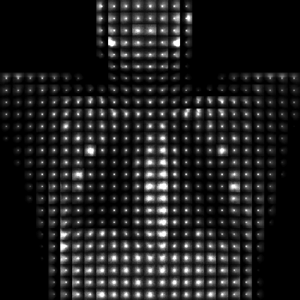
Image processing algorithms for compensation of the scattered radiation influence in X-ray imaging are proposed, studied and optimized by numerical simulation. These algorithms include the scattering estimation by convolution (superposition) technique, estimation of kernel functions by Monte Carlo (MC) simulation, the determination of the optimal number and shape of kernel functions and image segmentation. The determination of the number and shape of kernel functions was performed by the MC simulation of the realistic Zubal phantom and the clustering analysis of shape features of kernel functions. Testing simulation study of the algorithms for chest images at 75 keV proves that the optimal number of kernel functions is equal to 8. This number provides the three-fold contrast enhancement without using the anti-scatter grids. The achieved contrast is about 95% of the primary image contrast that exceeds contrast enhancements achieved with anti-scatter grids. An increased number of used kernel functions provides a better image contrast and better resolution of scattered radiation image, but estimation errors also increase due to the segmentation and deconvolution errors.References
- Z. Song, A. M. Fendrick, D. G. Safran, B. E. Landon, M. E. Chernew, “Global budgets and technology-intensive medical services,” Healthcare, vol. 1, no. 1–2, pp. 15–21, 2013, doi: https://doi.org/10.1016/j.hjdsi.2013.04.003.
- A. Assmus, “Early history of x rays,” Beam Line, vol. 25, no. 2, pp. 10–24, 1995, uri: https://www.slac.stanford.edu/pubs/beamline/25/2/25-2-assmus.pdf.
- M. J. Jensen, J. E. Wilhjelm, X-Ray Imaging: Fundamentals and Planar Imaging. Nutech: DTU, 2014.
- P. Monnin, F. R. Verdun, H. Bosmans, S. R. Pérez, N. W. Marshall, “A comprehensive model for x-ray projection imaging system efficiency and image quality characterization in the presence of scattered radiation,” Phys. Med. Biol., vol. 62, no. 14, pp. 5691–5722, 2017, doi: https://doi.org/10.1088/1361-6560/aa75bc.
- M. V. Kononov, O. A. Nagulyak, A. V. Netreba, “Influence of x-radiation in receiver system on reconstruction performance of projection tomography,” Radioelectron. Commun. Syst., vol. 51, no. 3, pp. 163–165, 2008, doi: https://doi.org/10.3103/S0735272708030084.
- S. Webb, Webb’s Physics of Medical Imaging, 2nd ed. Boca Raton: CRC Press, 2012, uri: https://www.routledge.com/Webbs-Physics-of-Medical-Imaging/Flower/p/book/9780750305730.
- I. Šabič, D. Ključevšek, M. Thaler, D. Žontar, “The effect of anti-scatter grid on radiation dose in chest radiography in children,” Cent. Eur. J. Paediatr., vol. 12, no. 1, pp. 75–80, 2016, uri: http://cejpaediatrics.com/index.php/cejp/article/view/273/pdf.
- E.-P. Rührnschopf, K. Klingenbeck, “A general framework and review of scatter correction methods in cone beam ct. part 2: scatter estimation approaches,” Med. Phys., vol. 38, no. 9, pp. 5186–5199, 2011, doi: https://doi.org/10.1118/1.3589140.
- W. Zhao, S. Brunner, K. Niu, S. Schafer, K. Royalty, G.-H. Chen, “A patient-specific scatter artifacts correction method,” in Progress in Biomedical Optics and Imaging - Proceedings of SPIE, 2014, vol. 9033, p. 903310, doi: https://doi.org/10.1117/12.2043923.
- P. G. F. Watson, E. Mainegra-Hing, N. Tomic, J. Seuntjens, “Implementation of an efficient monte carlo calculation for cbct scatter correction: phantom study,” J. Appl. Clin. Med. Phys., vol. 16, no. 4, pp. 216–227, 2015, doi: https://doi.org/10.1120/jacmp.v16i4.5393.
- K. Kim et al., “Fully iterative scatter corrected digital breast tomosynthesis using gpu-based fast monte carlo simulation and composition ratio update,” Med. Phys., vol. 42, no. 9, pp. 5342–5355, 2015, doi: https://doi.org/10.1118/1.4928139.
- A. V. Netreba, S. P. Radchenko, M. O. Razdabara, “Correlation reconstructed spine and time relaxation spatial distribution of atomic systems in mri,” in 2014 IEEE 34th International Scientific Conference on Electronics and Nanotechnology (ELNANO), 2014, pp. 365–367, doi: https://doi.org/10.1109/ELNANO.2014.6873453.
- Y. Suleimanov et al., “Magnetic resonance signal processing tool for diagnostic classification,” in 2016 IEEE 36th International Conference on Electronics and Nanotechnology (ELNANO), 2016, pp. 175–179, doi: https://doi.org/10.1109/ELNANO.2016.7493042.
- J. Maier, S. Sawall, M. Kachelriess, Y. Berker, “Deep scatter estimation (dse): feasibility of using a deep convolutional neural network for real-time x-ray scatter prediction in cone-beam ct,” in Medical Imaging 2018: Physics of Medical Imaging, 2018, vol. 10573, p. 56, doi: https://doi.org/10.1117/12.2292919.
- A. Y. Danyk, S. P. Radchenko, O. O. Sudakov, “Optimization of grid-less scattering compensation in x-ray imaging: simulation study,” in 2017 IEEE 37th International Conference on Electronics and Nanotechnology (ELNANO), 2017, pp. 316–320, doi: https://doi.org/10.1109/ELNANO.2017.7939770.
- A. Danyk, S. Radchenko, A. Netreba, O. Sudakov, “Using clustering analysis for determination of scattering kernels in x-ray imaging,” in 2019 10th IEEE International Conference on Intelligent Data Acquisition and Advanced Computing Systems: Technology and Applications (IDAACS), 2019, vol. 1, pp. 211–215, doi: https://doi.org/10.1109/IDAACS.2019.8924353.
- E. D. Prilepsky, J. E. Prilepsky, “Estimation of optimal parameter of regularization of signal recovery,” Radioelectron. Commun. Syst., vol. 61, no. 9, pp. 406–418, 2018, doi: https://doi.org/10.3103/S0735272718090030.
- I. A. Sushko, A. I. Rybin, “Speeding up the tikhonov regularization iterative procedure in solving the inverse problem of electrical impedance tomography,” Radioelectron. Commun. Syst., vol. 58, no. 9, pp. 426–433, 2015, doi: https://doi.org/10.3103/S0735272715090058.
- E.-P. Rührnschopf, K. Klingenbeck, “A general framework and review of scatter correction methods in x-ray cone-beam computerized tomography. part 1: scatter compensation approaches,” Med. Phys., vol. 38, no. 7, pp. 4296–4311, 2011, doi: https://doi.org/10.1118/1.3599033.
- I. G. Zubal, C. R. Harrell, E. O. Smith, Z. Rattner, G. Gindi, P. B. Hoffer, “Computerized three-dimensional segmented human anatomy,” Med. Phys., vol. 21, no. 2, pp. 299–302, 1994, doi: https://doi.org/10.1118/1.597290.
- D. Sarrut et al., “A review of the use and potential of the gate monte carlo simulation code for radiation therapy and dosimetry applications,” Med. Phys., vol. 41, no. 6Part1, p. 064301, 2014, doi: https://doi.org/10.1118/1.4871617.
- O. Sudakov, M. Kononov, I. Sliusar, A. Salnikov, “User clients for working with medical images in ukrainian grid infrastructure,” in 2013 IEEE 7th International Conference on Intelligent Data Acquisition and Advanced Computing Systems (IDAACS), 2013, vol. 2, pp. 705–709, doi: https://doi.org/10.1109/IDAACS.2013.6663016.
- L. Scrucca, M. Fop, T. B. Murphy, A. E. Raftery, “Mclust 5: clustering, classification and density estimation using gaussian finite mixture models,” R J., vol. 8, no. 1, p. 289, 2016, doi: https://doi.org/10.32614/RJ-2016-021.

Downloads
Published
2020-08-21
Issue
Section
Research Articles

